Iatrogenic left main coronary artery dissection: mind the catheter tip
Management of a potentially life-threatening complication of invasive coronary procedures

Iatrogenic left main coronary artery dissection: mind the catheter tip

Case Report
Issue
2016/10
DOI:
https://doi.org/10.4414/cvm.2016.00436
Cardiovascular Medicine. 2016;19(10):264-271

Affiliations
Department of Cardiology, Limassol General Hospital, Limassol, Cyprus

Published on 19.10.2016

Iatrogenic left main coronary artery (LMCA) dissection is a rare but dreadful complication of invasive coronary procedures with a reported incidence of <0.1% [1–4]. It is the result of mechanical trauma to the arterial wall during coronary artery instrumentation or man­ipulation leading to separation of the media by haemorrhage that creates a false lumen, with or without an associated intimal tear. The clinical presentation of iatro­genic LMCA dissection ranges from an asymptomatic, localised dissection with preserved blood flow to an extensive dissection leading to abrupt vessel closure and circulatory collapse. Timely recognition of the dissection and construction of a proper treatment plan based on the type of the dissection and the clinical status of the patient is needed to overcome this potentially fatal complication. Treatment consists of conservative therapy, salvage percutaneous coronary intervention (PCI) or urgent coronary artery bypass graft (CABG) surgery. Currently, iatrogenic LMCA dissection is most frequently treated with PCI, which has high procedural success and favourable long-term outcome [1–4]. Herein, we report two cases of catheter-­induced LMCA dissection of type C according to the National Heart, Lung and Blood Institute (NHLBI) criteria, which is considered a detrimental major type posing a high risk of adverse repercussions such as acute vessel closure [5–7]. Both patients were managed successfully with drug-eluting stent (DES)-facilitated PCI.

Case 1

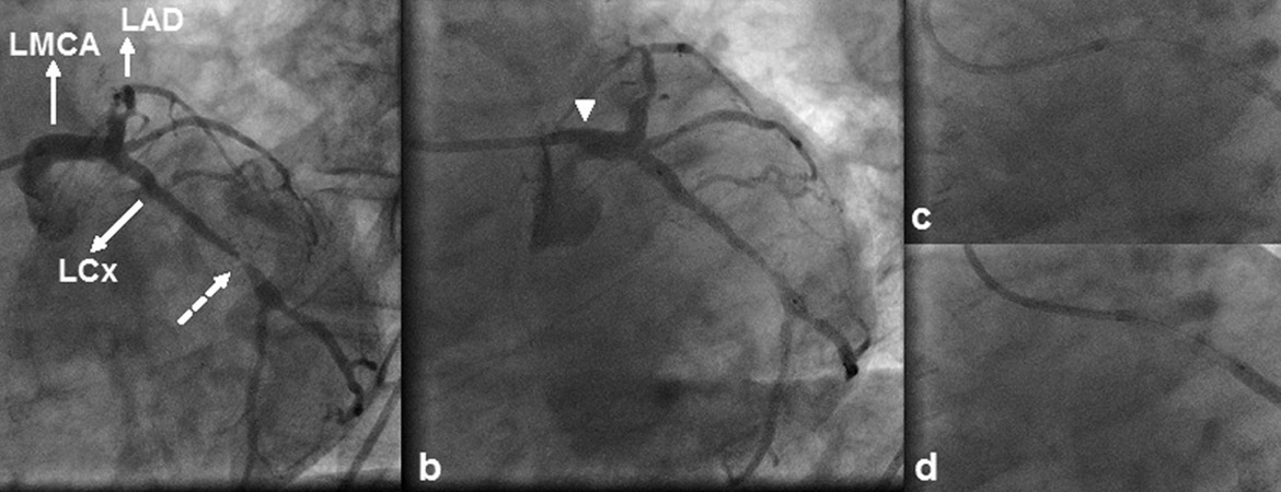
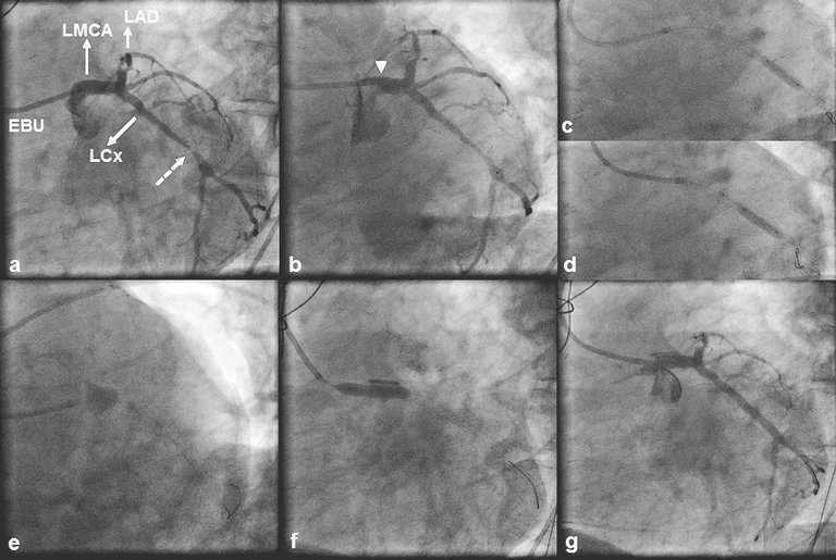
A 71-year-old, male patient was referred for coronary angiography because of non-ST-segment elevation myo­cardial infarction. The echocardiogram performed at the referring hospital showed hypokinesia of the inferior, inferior-septal and lateral left ventricular walls with an ejection fraction of 40%. The patient had a history of hypertension, hyperlipidaemia, inferior myocardial infarction and bare metal stent-facilitated PCI of a dominant right coronary artery (RCA) and the proximal and distal left circumflex (LCx) ­artery. In 2011, he underwent CABG with a left internal mammary artery graft to the left anterior descending (LAD) artery and a saphenous vein graft to a diagonal artery; preoperatively, no significant viability was documented with low dose dobutamine echocardiography over the dependent myocardium of the chronically occluded, yet collateralised RCA demonstrated during angiography. Transfemoral angiography during the current admission revealed patent grafts and obstructive in-stent disease of the proximal LCx artery culminating in a tight lesion just distal to the outflow of the stent (fig. 1a). Therefore, we proceeded with PCI to the LCx ­artery. The LMCA was engaged without difficulty with a 6 French Extra Back-up (EBU) 4.0 guiding catheter and, after predilation, a 3.5 × 33 mm DES was uneventfully deployed across the lesion (fig. 1b). ­Because of stent underexpansion at the site of the tight lesion we successfully performed postdilation with use of a 3.75 × 15 mm noncompliant balloon (fig. 1c). A second postdilation was then performed at a more proximal location (fig. 1d), yet subsequent angiography revealed persistent contrast staining outside the coronary lumen at the site of the LMCA ostium, which was compatible with a type C coronary dissection (fig. 1e). The patient was pain free and haemodynamically stable without electrocardiographic evidence of ­ischaemia. The dissection was immediately tackled with a 4.5 × 18 mm DES (fig. 1f). Postdilation was carried out with a 5.0 × 15 mm noncompliant balloon and final angiography showed complete sealing of the dissection flap (fig. 1g). Postprocedural creatine kinase and creatine kinase-MB isoenzyme levels were normal. The patient had an uneventful 2-day hospital course and was discharged home on life-long dual antiplatelet therapy. He remained stable 3.5 years post stenting with Canadian angina class I and no evidence of a cardio­vascular event.
Figure 1: (a) 45° left anterior oblique (LAO) view of the left coronary artery displaying a tight left circumflex (LCx) artery lesion (dashed arrow) located just distal to a previously implanted bare metal stent. A 6 French Extra Back-up (EBU) 4 guiding catheter is seen engaged in the ostium of a minimally diseased left main coronary artery (LMCA). The left anterior descending (LAD) artery, which contains a mid segment occlusion, is also shown. (b) LAO angiogram showing the stent deployment position. Note the unfavourable position of the guiding catheter resulting in the tip abutting against the wall of the LMCA ostium (arrow). Compared with its position during the first postdilation (c), the tip of the guiding catheter was too deep-seated during the second postdilation (d). (e) 40° LAO and 40° caudal view depicting persistent extraluminal contrast staining at the site of the LMCA ostium (type C coronary dissection). (f) Stent deployment across the dissection. (g) 45° LAO view showing an optimal angiographic result with complete sealing of the dissection flap.

Case 2

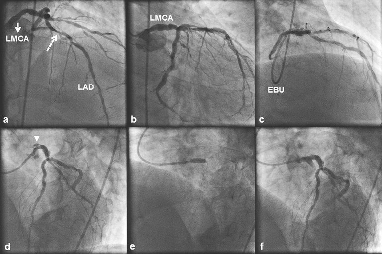
A 58-year-old male patient with a history of hyper­lipidaemia and cigarette smoking underwent transfemoral coronary angiography because of stable ­angina and ischaemia over the LAD artery territory, demonstrated with dobutamine stress echocardio­graphy. Angiography showed diffuse nonobstructive LMCA disease (fig. 2a, b), significant proximal LAD disease (fig. 2a) and chronic occlusion of a left posterior descending artery with faint filling through bridging collaterals. Therefore, we proceeded with PCI to the LAD artery lesion. The LMCA was engaged with a 6 French EBU 4.0 guiding catheter, and a 3.5 × 13 mm DES was directly implanted across the lesion (fig. 2c). Shortly thereafter the patient complained of acute, ­severe chest pain, and after multiple views focal and persistent extraluminal contrast staining that was compatible with a type C ostial LMCA dissection (fig. 2d) was revealed. The dissection was directly stented with a 4.0 × 15 mm DES (fig. 2e). Stent postdilation was performed with a 4.5 × 15 mm noncompliant balloon with a good final angiographic result (fig. 2f). Postprocedural creatine ­kinase and creatine kinase-MB isoenzyme levels were normal, and the patient was discharged home after a 2-day uneventful hospital course. He was prescribed life-long dual antiplatelet therapy. He remained stable 3.0 years after stenting with Canadian angina class I and no evidence of a cardiovascular event.
Figure 2: (a) 10° right anterior oblique (RAO) 40° cranial view of the left coronary artery displaying significant proximal left anterior descending (LAD) artery disease (dashed arrow). The left main coronary artery (LMCA) (a,b) contained substantial atheroma but no significant stenosis. (c) 40° RAO 40° cranial view obtained through a 6 French Extra Back-up (EBU) 4 guiding catheter showing the LAD artery stent deployment position. (d) 40° left anterior oblique (LAO) 20° cranial view after stenting displaying contrast outside the coronary lumen (“extraluminal cap”) at the LMCA ostium (arrowhead), which persisted after contrast had cleared from the coronary lumen (type C coronary dissection). (e) Stent deployment across the dissection. 
(f) 40° LAO 20° cranial view showing an optimal angiographic result with complete sealing of the dissection flap.

Discussion

Iatrogenic coronary artery dissection constitutes a complication with a significant impact on morbidity and mortality of patients undergoing diagnostic coronary angiography or PCI [8, 9]. In a multicentre study of 211 645 diagnostic cardiac catheterisations in the 1990s, the incidence of coronary artery dissection was 0.034% (71 cases) with a mortality of 0.0028% (6 cases) [10]. As shown in a large prospective PCI registry of nearly 21 000 patients, the incidence of in-laboratory severe coronary artery dissection (NHLBI dissection type ≥C or abrupt closure) decreased over the years from 1.0% in the prestent era to 0.7% in the first-generation stent era to 0.3% in the contemporary stent era (2000–2003) [11]. However, in contemporary practice, severe coronary artery dissection accounted for 6.2% of all PCI failures and was the most common reason (80%) for referring patients for emergency CABG after failed PCI. Coronary artery dissections may be caused by several mechanisms. Mechanical dilation of a coronary artery by ­angioplasty balloon inflation or stent implantation is associated with mechanical trauma to the vessel wall, which is a function of the biomechanical properties of the plaque and is the basis of an inherent risk of these procedures – coronary artery dissection. Accordingly, calcified, eccentric and long lesions, “complicated” lesions (ulcerated, thrombus-laden) and ­lesions located in angulated coronary segments carry a higher risk for the development of dissection [5]. Technical factors increasing the risk of iatrogenic coronary ­artery dissection include the use of stiff-tipped or hydrophilic-tipped guidewires to cross tightly narrowed or totally occluded arteries, angioplasty balloon overinflation or oversizing (balloon to artery ratio >1.2), not coaxially and/or deeply engaged catheters, large-bore catheters and Amplatz-shaped catheters [12, 13]. Reported risk factors for catheter-induced coronary ­artery dissection include atherosclerotic disease, catheterisation for acute myocardial infarction, and variant anatomy of the coronary ostia necessitating extensive catheter manipulations, vigorous contrast media injection, and vigorous, deep inspiration [12]. As far as the arterial access site for performing coronary catheterisation is concerned (transfemoral versus trans­radial approach), no difference with regards to non-­access site complications, including coronary artery dissection [14], has been reported. Nonetheless, a “universal” catheter, that is, a catheter that can be used for left and right transradial diagnostic coronary angio­graphy and/or PCI, such as the Kimny catheter, may be associated with an increased risk of coronary artery dissection because of a difficult coaxial engagement or deep engagement, especially in a RCA with an inferior takeoff or when the catheter is removed from the LMCA without initial downward pressure and torque [2, 15]. Despite the decrease in the incidence of iatrogenic coronary artery dissection observed over the years, the constantly increasing complexity of PCIs (use of large-bore catheters and stiff-tipped guidewires for recanalisation of chronically occluded arteries, retro­grade PCI, PCI to the LMCA, PCI to calcified bifurcation lesions) render iatrogenic coronary artery dis­section a meaningful risk mandating good comprehension of its mechanisms and predisposing factors, as well as its angiographic presentation and management.
The RCA is the most frequently dissected vessel (84–87% of the cases), followed by the LAD, left main coronary and LCx arteries [16, 17]. Iatrogenic dissection of both the RCA and LMCA is mostly observed after inadequate alignment of a diagnostic or guiding catheter (1,2,4), yet the LMCA and RCA arise from their respective aortic sinuses at different angles: acute (range 20°–55°) and almost perpendicular (range 60°–88°), respectively. This might render the LMCA less susceptible to catheter-induced dissection by providing a better ­approach for catheterisation [17]. Three retrospective case series have reported the incidence of iatrogenic LMCA dissection during a coronary catheterisation procedure (PCI or diagnostic coronary angiography). Lee et al. [1] reported an incidence of 0.03% (10 cases out of 34 190 procedures), Cheng et al. [2] an incidence of 0.071% (13 cases out of 18 400 procedures), and ­Eshtehardi et al. [4] an incidence of 0.07% (38 cases out of 51 452 procedures) with a twofold greater incidence of iatrogenic LMCA dissection during PCI (0.1% of all PCIs) than during diagnostic coronary angiography (0.06% of all diagnostic coronary angiographies). Dissection of the LMCA is most frequently caused by inappropriate positioning of the diagnostic or guiding catheter, with an incidence of 61.5% in the study by Cheng et al. [2] where a 6 French catheter was used in 84.6% of the cases, the Kimny miniradial catheter in 61.5% of the cases and the left Judkins catheter in 30.8% of the cases. Balloon dilation near the LMCA bifurcation and stenting at the LAD artery ostium were the second and third most frequent causes of LMCA dissection in this study, with an incidences of 23.1% and 7.7%, respectively. In the study by ­Eshtehardi et al. [4], inappropriate positioning of a diagnostic catheter was implicated in 58% of the cases of LMCA dissection where the left Judkins catheter was used in 82% of the cases; inappropriate positioning of a guiding catheter was implicated in 16% of the cases of LMCA dissection where extra backup catheters (Amplatz left-, EBU- or Q-curve) were used in 56% of the cases. The second most frequent cause of LMCA dissection in this study was deep intubation of the guiding catheter during balloon retrieval, which was observed in 26% of the cases. In our first case, as shown in fig. 1b, the 6 French EBU 4.0 guiding catheter was non-coaxially positioned and its tip abutted against the wall of the LMCA ostium without, however, causing pressure damping or ventricularisation. However, such a catheter position suggested that the catheter might have been “too short” for the patient and that a more coaxial LMCA engagement could have been achieved with an EBU 4.5 guiding catheter. Dissection of the LMCA occurred secondary to deep seeding of the guiding catheter and scraping of the LMCA wall during retrieval of the postdilating balloon. If we had disengaged the guiding catheter from the LMCA and pulling, to keep the catheter out of the LMCA ostium, had been more vigorous to withstand the resistance met during retrieval of the postdilating balloon, we would have prevented deep seeding of the guiding catheter and the resultant LMCA dissection. In our second case, the LMCA contained substantial, but nonobstructive atheroma. As shown in fig. 1d, the 6 French EBU 4.0 guiding catheter was non-coaxially positioned with its tip pointing ­vertically against the roof of the LMCA. Accordingly, a ­hydraulic LMCA dissection might have been created during contrast injection. Keeping the catheter coaxially positioned during every minute of the procedure, avoiding contrast media injection in the presence of pressure damping or ventricularisation and gradual ramping of the injection are essential actions in order to minimise the risk of LMCA dissection.
Iatrogenic dissection of the ascending aorta during cardiac catheterisation procedures is a rare complication and mainly a sequela of coronary artery dissection extending in a retrograde fashion into the aortic root; it is significantly more likely to occur during PCI than during a diagnostic procedure, with incidences ranging from 0.07% to 0.6% and from 0.01% to 0.08%, respectively [18]. The right aortic sinus is involved in more than 50% of the cases, suggesting that it may be vulnerable to retrograde extension of an RCA dissection [17–19]. The periostial aortic wall of the RCA has less interstitial type I collagen than the periosteal aortic wall of the LMCA. Also, the sinotubular ridge in the right aortic sinus has a smaller amount of smooth muscle cells within an extracellular matrix basically composed of type III collagen, whereas the sinotubular ridge in the left aortic sinus has a larger amount of smooth muscle cells within a dense extracellular ­matrix of type I collagen [17]. Because the tensile strength of type I collagen is greater than that of type III collagen, the RCA might thereby have less resistance to traction and RCA dissection might more easily extend retrogradely to involve the aortic root . In a retrospective series of 18 patients with iatrogenic aortic ­dissection occurring during cardiac catheterisation procedures reported by Gómez-Moreno et al. [18], the dissection involved the RCA and its corresponding ­aortic ­sinus in 67% of the cases, and was most often ­related to deep coronary catheter engagement and the use of unconventional catheters (Amplatz, XB, multipurpose) (39%). Balloon dilation, crossing of chronic ­total occlusion with a guiding wire and stent implantation were identified as additional causes of the dissection. Núñez-Gil et al. [19], in their retrospective series of 74 patients with iatrogenic aortic dissection occurring during cardiac catheterisation procedures found that the dissection, in the vast majority of cases (97.2%), took place during coronary catheter engagement of the RCA (56.8%) or the LMCA (40.5%) and was caused by a catheter (91.8%) of 6 French size (90.5%). Unconventional catheters (Amplatz, XB, multipurpose) were used in 48.6% of the cases and guiding catheters were used in 70.3% of the cases.
There are several precautionary measures that can be taken in order to minimise the risk of catheter-induced coronary artery dissection. The first is the optimal ­selection of the catheter with adequate coaxial engagement of this catheter into the coronary artery followed by careful catheter handling. For left coronary artery catheterisation from the transfemoral approach in ­patients with a normal-sized aortic root and a normal length of the LMCA, the Judkins left 4.0 and 4.0 extra back-up type guiding catheter are good choices, downsizing to 3.5 or upsizing to 4.5 as needed [20]. Catheter-induced dissection of the roof of the LMCA in ­patients with a large-sized aortic root is commonly ­observed when using a “too short” Judkins left 4.0 or 3.5 backup type guiding catheter. In patients with a large-sized aortic root the Judkins left 5.0 or extra backup type 4.0–4.5 guiding catheter are good choices. Compared with the transfemoral approach, left coronary artery catheterisation from the right transradial approach in patients with a normal-sized aortic root is usually performed with catheters having a 0.5 cm shorter curve (Judkins left 3.5 and extra backup type 3.5 guiding catheter). If possible, catheter engagement in a coronary ostium must be performed with the catheter connected to continuous pressure monitoring in order to ensure that there is no pressure damping or ventricularisation, thereby avoiding inadvertent dissection during contrast injection. Gradual ramping of the injection can also help to minimise the risk of this event. Given that the extra backup type guiding catheters have been implicated in iatrogenic LMCA dissection, such catheters should be selected only in cases of complex PCI requiring a strong backup support. Maintaining coaxial alignment of the guiding catheter with the coronary ostium during the passage of interventional devices (stents, conventional balloons, cutting balloons, rotational ablative or distal protection ­devices, etc.) is important because these devices are usually rigid and of large profile and their passage through a non-coaxially engaged guiding catheter may lead to ostial dissection. Deep seating of the guiding catheter in order to achieve a strong “active backup” must be performed with extreme care and when the catheter tip is soft, if the artery is large enough to accommodate the catheter and there is no ostial or proximal lesion. Also, the guiding catheter must be first disengaged from the ostium and be kept there by continuous pulling during ­retrieval of interventional devices, in order to avoid coronary dissection secondary to deep seating of the guiding catheter. Extreme care is also required when using Amplatz-shaped catheters, since a simple withdrawal from the vessel can cause the tip to advance further into the vessel and cause dissection. In order to disengage the Amplatz catheter, one must first advance it under fluoroscopy to prolapse the tip out of the ostium and then rotate it so that the tip is totally out of the ostium before withdrawing it.
Angiographically, coronary dissection appears as a radio­lucent area within the vessel or as an extravasation of contrast agent. Based on their angiographic ­appearance, coronary dissections are classified into six types (type A to F), according to the NHLBI classification scheme [5]. As shown in studies conducted in the angioplasty era, the angiographic morphology of the dissection is associated with the clinical outcome, and it can thus help in selection of the most appropriate treatment strategy [6]. Dissection types C to F (type C: contrast appears outside the coronary lumen as an “extraluminal cap” with persistent contrast staining; type D: spiral luminal filling defects, often with persistent contrast staining; type E: new, persistent intra­luminal filling defects; and type F: dissection without any of the morphological characteristics described in this classification that is associated with impaired flow or total coronary occlusion) are characterised as major dissections having a significant risk of in-hospital complications such as acute vessel closure (31%), need for emergency CABG (37%), myocardial infarction (13%) and repeat angioplasty (24%) [5, 6]. In contrast, dissection types A and B (type A: minor intraluminal radio­ucent areas with minimal or no persistent contrast staining, and type B: radiolucent tracks representing the luminal flap and coursing parallel to the vessel or a double lumen appearance separated by the radiolucent luminal flap with minimal or no persistent contrast staining) have not been shown to increase morbidity and mortality compared with those of patients without dissection, and neither do they affect procedural outcome; the incidence of abrupt vessel closure, myocardial infarction and need for CABG in patients with type B dissections has been reported to be less than 3% [6]. Alternatively, LMCA dissection can be classified into three types according to a simplified classification scheme described by Eshtehardi et al. [4]. Type I dissections are localised and do not extend into the LAD or LCx arteries, type II dissections are characterised by extension into the LAD and LCx arteries, and type III dissections are those extending back to involve the aortic root. Angiographically, iatrogenic aortic dissection appears as dense and persistent contrast staining of the aortic wall. A classification scheme proposed by Dunning et al. [21] recognises three classes of iatrogenic aortic dissection based on the extent of aortic involvement in the dissection. Class I dissections are limited to the corresponding aortic sinus, Class II dissections involve the corresponding aortic sinus and extend less than 40 mm into the aorta and Class III dissections involve the corresponding aortic sinus and extend more than 40 mm into the aorta. Depending on whether anterograde flow has been impaired and to what degree, the clinical spectrum of LMCA dissection ranges from an asymptomatic status to refractory cardio­genic shock and/or cardiac arrest. In the series of 38 patients with iatrogenic LMCA dissection reported by Eshtehardi et al. [4], no patient with type I dissection (21 patients) manifested haemodynamic instability. In contrast, 7 (41%) of 17 patients with type II or III dissections presented ­haemodynamic instability and 5 of these patients (29%) required cardiopulmonary resuscitation. Both our patients were diagnosed with a type C dissection according to the NHLBI classification scheme. Also, in both our patients, the LMCA dissection was localised and did not extend to the LAD or LCx arteries or retrogradely into the aortic root, thereby qualifying as a type I dissection according to the simplified classification scheme of Eshtehardi et al. [4]. Antero­grade blood flow and haemodynamic stability were maintained in both cases.
Iatrogenic LMCA dissection is an emergency because it threatens a large territory downstream of the injury, and its management depends on the patency of the distal vessel and the extent of propagation of the dissection. Percutaneous or surgical revascularisation is generally mandated in the presence of myocardial ­ischaemia or acute vessel closure, whereas conservative management has been advocated in asymptomatic and haemodynamically stable patients with local­ised dissections and normal distal coronary flow. The currently prevailing management strategy of iatro­genic LMCA dissection that produces ischaemia is PCI, which can be performed rapidly after the occurrence of dissection with a high technical success rate and acceptable short- and long-term outcomes. PCI circum­vents the delays associated with CABG and can expeditiously restore of coronary patency, thereby avoiding prolonged ischaemia, which is linked to an increased rate of myocardial infarction and death, something that is particularly important for the haemo­dynamically unstable patient. A literature ­review of bail-out PCI for iatrogenic LMCA dissection that included 54 patients revealed a procedural success rate of 92.6%, whereas only four patients underwent emergent CABG as a result of unsuccessful PCI [3]. The overall survival rate was 92.6% and of the four deaths recorded only two were of cardiac origin. In the series by Eshtehardi et al. [4], there was a 37% (14/37) rate of bail-out PCI and a 45% (17/37) rate of emergency CABG without in-hospital mortality, whereas at 5 years no significant difference was observed between the two revascularisation strategies with regards to major ­adverse cardiac events (36% vs 41%, respectively; p = 0.8). Surgical revascularisation is reserved for patients in whom PCI failed to treat LMCA dissection or for haemo­dynamically stable patients who otherwise would have been deemed surgical candidates on the basis of extensive multivessel coronary disease. Since surgery does not treat the dissection itself, surgical revascularisation solely for LMCA dissection not producing significant lumen compromise is not an appropriate treatment strategy, because of the risk of graft closure. Eshtehardi et al. [4], also reported that 6 of their 37 patients (16%) had a localised and stable LMCA dissection and received conservative treatment with favourable short- and long-term outcomes. Furthermore, in 12 of their patients (32%), the LMCA dissection showed signs of expansion within 90 minutes of the initial observation period, something that highlights the dynamic nature of the dissection and its potential to rapidly transform into an extensive dissection leading to haemodynamic collapse due to abrupt flow compromise with disastrous sequelae. However, Eshtehardi et al. [4] did not provide any information about the type(s) of dissection that expanded. Accordingly, when confronted with a patient with an iatrogenic LMCA dissection, the decision to intervene with either PCI or CABG or to treat the patient medically must take into account whether the dissection is minor or major based on several angiographic signs, the clinical status of the patient, the operator’s expertise and availability of equipment (intravascular imaging systems) to perform PCI to the LMCA successfully, and the time ­required to transfer the patient for CABG. Whilst major LMCA dissections (the dissections extending more than 20 mm, causing at least 50% residual stenosis and impairing flow, as well as dissection types C to F ­according to NHLBI criteria) generally require prompt PCI, dissection types A and B can be treated conservatively and under close monitoring with optimal medical therapy including a β-blocker. Conservative therapy has been applied successfully in highly selected patients with minor and asymptomatic LMCA dissections, yet late progression of an initially localised LMCA dissection into an expanding false lumen leading to significant reduction in luminal diameter with associated exertional angina has been described [22, 23]. The decision to treat a patient with LMCA dissection medically must therefore be accompanied by a revascularisation plan.
Both our patients were haemodynamically stable and had a type C LMCA dissection, which is considered a major type with a 10% risk of acute vessel closure [6, 7]. Acute LMCA occlusion is usually associated with rapid haemodynamic deterioration and cardiac arrest, but in such an unwanted event, our first patient would have faced less risk of haemodynamic compromise than our second patient owing to the presence of patent grafts to the LAD and diagonal arteries; however, the resultant significant ischaemia in the LCx artery territory could still cause some haemodynamic compromise because of his moderately reduced ejection fraction (40%) associated with his previous acute inferior myocardial infarction. Therefore, both patients were treated with implantation of a stent with complete dissection coverage, and both had a favourable immediate and long-term outcome. During PCI of a dissected coronary ­artery, insertion of a soft-tipped guiding wire into the true lumen is a crucial step of the procedure, and in the case of LCMA dissection both the LAD and LCx arteries should be wired in order protect them from possible extension of the dissection. Intracoronary imaging with means of intravascular ultrasound or optical coherence tomography can be very helpful if the position of the guiding wire is doubtfull, and it can also help define the dissection entry point and extent, the existence and extent of intramural haematoma, and vessel size, thereby facilitating PCI [24]. Intracoronary imaging for PCI of a dissected coronary artery ensures adequate stent coverage of the whole dissection flap, thereby preventing dissection/haematoma propagation that could result from inadvertent premature sealing of the dissection flap. Alternatively, conservative treatment with stenting of the dissection entry site only may be sufficient to stabilise this complication, providing that the residual false lumen is not obstructive and normal anterograde flow is obtained. This approach has been applied in a case of LMCA dissection reported by Binder et al. [25]. During PCI to a calcified proximal LAD artery lesion, the predilating balloon ruptured and produced an ostial LAD artery dissection extending retrogradely into the LCMA. They delivered two overlapping stents in the proximal and ostial LAD artery followed by examination of the LMCA and the LAD artery by means of optical coherence tomography. They found that the dissection entry site was located in the ostial LAD artery and was adequately covered by the stent, whereas the false lumen did not cause obstruction to the LMCA. Therefore they refrained from stenting the LMCA and at 6-months angio­graphic follow-up, no evidence of residual LMCA dissection or stenosis was documented. Similarly, conservative treatment with stent implantation sealing the dissection entry site in the coronary artery has also been reported to be a successful approach in about 50% of the patients with retrograde extension of the dissection in the ascending aorta [18, 19].

Conclusion

Iatrogenic LMCA dissection is a rare and potentially life-threatening complication of invasive coronary procedures. The fact that iatrogenic LMCA dissection is mostly catheter-induced underlies the need for proper catheter selection on the basis of the patient’s anatomy and the complexity of the PCI, meticulous handling of coronary catheters with adequate coaxial engagement during every minute of the procedure, and gradual ramping of contrast media injection in the absence of pressure damping or ventricularisation. In the case of an iatrogenic LMCA dissection, prompt diagnosis and construction of a treatment plan is needed in order to overcome this potentially detrimental complication. Dissection types C to F according to NHLBI criteria are considered major dissections posing significant risk of adverse clinical outcomes if left untreated. Currently, bail-out PCI for iatrogenic LMCA dissection appears to be safe and feasible with acceptable short- and long-term outcomes. CABG is a valid treatment strategy in patients without haemodynamic instability who otherwise would have been deemed surgical candidates on the basis of extensive multivessel coronary disease. Conservative therapy may be considered in haemodynamically stable and clinically asymptomatic patients with localised dissections (types A and B according to NHLBI criteria).
No financial support and no other potential conflict of interest ­relevant to this article was reported.
Correspondence:
Andreas Y. Andreou, MD
Department of Cardiology
Limassol General Hospital
Nikeas street,
Pano Polemidia
Postal code 3304, PO Box
CY-56060, Limassol
 1 Lee SW, Hong MK, Kim YH, Park JH, Rhee KS, Lee CW, et al. Bail-out stenting for left main coronary artery dissection during catheter-based procedure: acute and long-term results.Clin Cardiol. 2004;27(7):393–5.
 2 Cheng CI, Wu CJ, Hsieh YK, Chen YH, Chen CJ, Chen SM, et al. Percutaneous coronary intervention for iatrogenic left main coronary artery dissection. Int J Cardiol. 2008;126(2):177–82.
 3 Onsea K, Kayaert P, Desmet W, Dubois CL. Iatrogenic left main ­coronary artery dissection. Neth Heart J 2011;19(4):192–5.
 4 Eshtehardi P, Adorjan P, Togni M, Tevaearai H, Vogel R, Seiler C,
et al. Iatrogenic left main coronary artery dissection: incidence, ­classification, management, and long-term follow-up. Am Heart J 2010;159(6):1147–53.
 5 Rogers JH, Lasala JM. Coronary artery dissection and perforation complicating percutaneous coronary intervention. J Invasive ­Cardiol.2004;16(9):493–9.
 6 Huber MS, Mooney JF, Madison J, Mooney MR. Use of a morphologic classification to predict clinical outcome after dissection from coronary angioplasty. Am J Cardiol.1991;68(5):467–71.
 7 Klein LW. Coronary complications of percutaneous coronary ­intervention: a practical approach to the management of abrupt closure. Catheter Cardiovasc Interv. 2005;64(3):395–401.
 8 de Bono D. Complications of diagnostic cardiac catheterisation: results from 34 041 patients in the United Kingdom confidential enquiry into cardiac catheter complications. The Joint Audit Committee of the British Cardiac Society and Royal College of Physicians of London.Br Heart J.1993;70(3):297–300.
 9 Holmes DR Jr, Holubkov R, Vlietstra RE, Kelsey SF, Reeder GS, ­Dorros G, et al. Comparison of complications during percutaneous transluminal coronary angioplasty from 1977 to 1981 and from 1985 to 1986: the National Heart, Lung, and Blood Institute Percutaneous Transluminal Coronary Angioplasty Registry. J Am Coll ­Cardiol. 1988;12(5):1149–55.
10 West R, Ellis G, Brooks N. Complications of diagnostic cardiac ­catheterisation: results from a confidential inquiry into cardiac catheter complications. Heart. 2006;92(6):810–4.
11 Mattichak SJ, Dixon SR, Shannon F, Boura JA, Safian RD. Failed percutaneous coronary intervention: a decade of experience in 21 000 patients. Catheter Cardiovasc Interv.2008;71(2):131–7.
12 Boyle AJ, Chan M, Dib J, Resar J. Catheter-induced coronary artery dissection: risk factors, prevention and management. J Invasive Cardiol.2006;18(10):500–3.
13 Ammann P, Brunner-La Rocca HP, Angehrn W, Roelli H, Sagmeister M, Rickli H. Procedural complications following diagnostic coronary angiography are related to the operator’s experience and the catheter size. Catheter Cardiovasc Interv.2003;59(1):13–8.
14 Hibbert B, Simard T, Wilson KR, Hawken S, Wells GA, Ramirez FD,
et al. Transradial versus transfemoral artery approach for coronary angiography and percutaneous coronary intervention in the extremely obese. JACC Cardiovasc Interv.2012;5(8):819–26.
15 Caputo RP, Tremmel JA, Rao S, Gilchrist IC, Pyne C, Pancholy S, et al. Transradial arterial access for coronary and peripheral procedures: executive summary by the Transradial Committee of the SCAI. Catheter Cardiovasc Interv. 2011;78(6):823–39.
16 Zidi M, Nallet O, Esteve JB, Michaud P, Cattan S. Extensive iatrogenic coronary dissection during coronary angioplasty: a series
of 19 consecutive patients. Ann Cardiol Angeiol (Paris). 2010;59(5):306–10.
17 López-Mínguez JR, Climent V, Yen-Ho S, González-Fernández R, Nogales-Asensio JM, Sánchez-Quintana D. Structural features of the sinus of valsalva and the proximal portion of the coronary arteries: their relevance to retrograde aortocoronary dissection. Rev Esp Cardiol.2006;59(7):696–702.
18 Gómez-Moreno S, Sabaté M, Jiménez-Quevedo P, Vázquez P, Alfonso F, Angiolillo DJ, et al. Iatrogenic dissection of the ascending aorta following heart catheterisation: incidence, management and outcome. EuroIntervention.2006;2(2):197–202.
19 Núñez-Gil IJ, Bautista D, Cerrato E, Salinas P, Varbella F, Omedè P, et al. Incidence, Management, and Immediate- and Long-Term Outcomes After Iatrogenic Aortic Dissection During Diagnostic or Interventional Coronary Procedures. Circulation 2015;131(24):2114–9.
20 Pavei A, Marco J. Tools & techniques: choice and use of guiding catheters. EuroIntervention.2010;6(4):543–4.
21 Dunning DW, Kahn JK, Hawkins ET, O’Neill WW. Iatrogenic coronary artery dissections extending into and involving the aortic root. Catheter Cardiovasc Interv.2000;51(4):387–93.
22 Murat C, Uygar CY, Emre Y, Yalcin G, Atila I. Conservative treatment of iatrogenic left main coronary artery dissection: report of two cases. Cardiovasc Diagn Ther. 2013;3(4):244–6.
23 Zelinger AB, Shulruff S, Pouget JM. Significant left main stenosis following asymptomatic dissection during coronary arteriography. Chest 1983;83(3):568–9.
24 Sanidas E, Buysschaert I, van Langenhove G. Iatrogenic left main coronary artery dissection and intramural hematoma caused by diagnostic transradial cardiac catheterization. Hellenic J
­Cardiol
.2014;55(1):65–9.
25 Binder RK, Boone RH, Webb JG. Left main dissection conservatively managed with optical coherence tomography guidance. Catheter Cardiovasc Interv.2014;83(1):65–8.