The evolution of surgical valves
Lessons can be learned from the history of heart valve prostheses

The evolution of surgical valves

Review Article
Issue
2017/12
DOI:
https://doi.org/10.4414/cvm.2017.00532
Cardiovascular Medicine. 2017;20(12):285-292

Affiliations
a University Heart Center, Universitätsspital Zürich, University of Zurich, Switzerland; b Cardio-Vascular Research Department of Surgery and ­Anesthesiology, CHUV, Lausanne, Switzerland

Published on 20.12.2017

Introduction

On 7 September 1896, a 22-year-old man was stabbed in the heart and collapsed. Two days later Dr Ludwig Rehn, from Frankfurt, performed the first reported heart surgery operation, suturing the wound in the heart through a left thoracotomy approach [1]. Since that time, many changes have occurred and cardiovascular surgery has evolved exponentially since its beginning in 1953, when, John Gibbon performed the first closure of an atrial septal defect with use of a heart-lung machine. But the history of surgical valve treatment starts even earlier. Before the availability of the heart-lung machine, valve surgery was performed via a closed approach, on the beating heart.
The first “closed heart procedure” was performed in 1914 when Theodor Tuffier treated an aortic valve stenosis by digitally opening the valve through the aortic wall [2]. In 1923, Elliot Carr Cutler, in conjunction with his cardiology colleague, Samuel Levine, performed a closed transventricular mitral commissurotomy.
Digital commissurotomy was introduced in 1948 by Bailey in Philadelphia and Harken in Boston and for many years was the treatment of choice for patients with mitral valve stenosis. The first valve prosthesis was a “sutureless valve”: Charles Hufnagel [3], in 1952, implanted an heterotopic valvular heart prosthesis in the descending aorta of a patient with aortic valve regurgitation. This represented the first step in a long journey that lasted more than 50 years and is not finished yet. It is inspiring that most modern-generation surgical valves have some features already researched in the early days of surgical evolution, such as ­sutureless implantation, plastic leaflets and the beating heart approach.
Heart valve innovation has been one of the most ­important factors influencing the evolution of cardiovascular medicine. Denton Cooley often said ­“Apply, Simplify, Modify”. This philosophy inspired generations of cardiac physicians and describes well what happened in the evolution of heart valve prostheses. Continuous and passionate research into new ­materials, technologies and techniques to overcome the infinite challenges of replacing a natural structure with an artificial implant.
Heart valve innovation is also a good example of team-work, between physicians and engineers. Albert Starr, a surgeon from Colombia University and Lowell ­Edwards, an engineer close to retirement, met in 1957 and created the first commercial mechanical valve prosthesis with a long history of successful implants: the Starr-Edwards balloon cage prosthesis. A multi­disciplinary team composed of a cardiac surgeon, Dr Nicoloff, an industrial engineer, Dr Posis, and an entrepreneur, Manuel Villafana, together developed, in 1976, the first bileaflet prosthesis [4].
The early days of prosthetic valve development provided much information that is still of value today. What can we learn from the good, the bad and the ugly experiences of the pioneers of valve innovation?
We would like to give an overview of the history and ­evolution of heart valves, focusing not only on the technical features of each prosthesis, but also on the trends and mechanisms that influenced this continuous development. We have divided the paper in sections that ­each describe the evolution of a subtype of heart valve, from mechanical and biological surgical prostheses to the transcatheter valves, and explain the reasons that ­determined the emergence of the “next-step valve”. With this purpose, we are not going to mention all the devices, but take example of the major ones to understand the trend of evolution.

First steps in heart valve surgery

The evolution of heart valves began in the late 1940s, when, Charles Hufnagel designed a methacrylate chamber containing a methacrylate ball that was ­implanted in the descending aorta of a patient with aortic regurgitation. More that 200 patients were treated after 1952 [5]. The opportunity to work with an open heart permitted Dwight Harken, in 1960, for the first time to implant, in an annular position, a “double-caged ball” prosthesis called the Harken-Soroff[6, 7]. In the same year, Nina Braunwald started her experience with mitral valve replacement using a flexible poly­urethane mitral prosthesis with attached Teflon chordae tendinae [8] and Albert Starr performed the first mitral valve replacement with the Starr-Edwards ball-valve. This valve was inspired by an old bottle stopper and was developed as a ball valve with a single methacrylate cage and a Silastic ball inside, as occluder.
The first results of this procedure were published in 1961 in an enthusiastic and innovative manuscript, which is still inspirational today. A careful reading of the original paper of Albert Starr and Lowell Edwards reveals challenges and questions that are still valid today, and that affected the evolution of the last generation of surgical valves, endovascular implantable transcatheter valves [9]. The authors were confronted with the (still) difficult choice between the more physio­logical option of valve repair and the more reproducible and reliable option of valve replacement.
A high operative mortality represented the first limiting factor, mostly related to the complexity of operation and perioperative care. A lower profile valve that enabled easier and faster implantation appeared to be mandatory from the first. This was one of the first steps in the evolution of heart valve design: Starr modified the Harken valve by removing the second cage to simplify implantation. The issue of durability was raised as long ago as 1961. The Starr-Edwards valve was tested in vitro and, according to the results, a durability of 40 years would have been expected. Recently this hypothesis was confirmed [10].
The haemodynamic performance of the heart prosthesis should be as close as possible to the native “perfect” valve, with low resistance to the forward flow and ­allowing only trivial regurgitant backflow once the ­occluder closes [11]. Paravalvular leakage and the risk of endocarditis were immediately detected: they were the indications for ­reoperation in the two surviving patients of Harken’s initial series. Valve noise was recognised as an important problem and was solved early, for instance by replacing the methacrylate ball in the Hufnagel prosthesis with a nylon one coated with a ­silicone rubber. Initial preclinical studies with mechanical valves showed the high level of anticoagulation needed to avoid valve occlusion and an elevated risk of thromboembolic events was also described.
All these aspects were already clear in the first decades of heart valve surgery and they have steadily guided the evolution of heart valve prostheses. These “old concepts” will be the “key words” adopted in this review, to explain the prostheses’ evolution.

Mechanical heart valves: past, ­present and future

The poor haemodynamic performances of the “ball-cage” valves indicated a need for the development of a second generation of mechanical prostheses. In fact, the central ball occluder caused lateralisation of ­forward flow and therefore high turbulence; moreover, the high profile and the large sewing ring produced a restricted effective orifice area (EOA), and limited efficacy in the mitral position, with the risk of outflow tract obstruction [11, 12].
The need for the central flow, reproducing a more physio­logical pattern, led, at the end of the 1960s, to the development of tilting-disk prostheses. The Björk-Shiley valve was the first tilting-disc prosthesis to be widely implanted: it was designed with a central disk hold in place by two struts [13]. The open valve had two orifices, with the turbulent flow limited to the area near to the occluder. The flow ­resistance was related to the disc design and to the ­degree of the opening angle, and for this reason the disc was progressively modified into a convexo-concave shape that could slide about 2 mm during its movement, increasing the EOA. These minor engineering modifications, with the aim to achieve a better haemodynamic profile, led unexpectedly to a higher incidence of leaflet blockade and ­embolisation due to the excessive “leverage-loading” on the outflow strut [13]. This brought about the end of production of this prosthesis. The history of the Björk-Shiley is a paradigmatic example of how delicate the evolution of mechanical heart valves was.
Hoping to improve haemodynamics, Kalke and ­Lillehei developed the first prototype of a rigid bileaflet valve, but very limited clinical use was reported. In 1977, the St. Jude Medical (SJM) bileaflet prosthesis was introduced and implanted by Nicoloff and associates [14]. This design produces three flow areas through the valve orifice, with a more uniform and laminar central flow. Better haemodynamics was associated with less blood stagnation and the lower profile allowed easier implantation. Recently, the valve has been redesigned as the SJM Regent valve. The sewing ring and the external profile were modified to further increase the effective orifice area, especially in the smaller aortic prostheses [15].
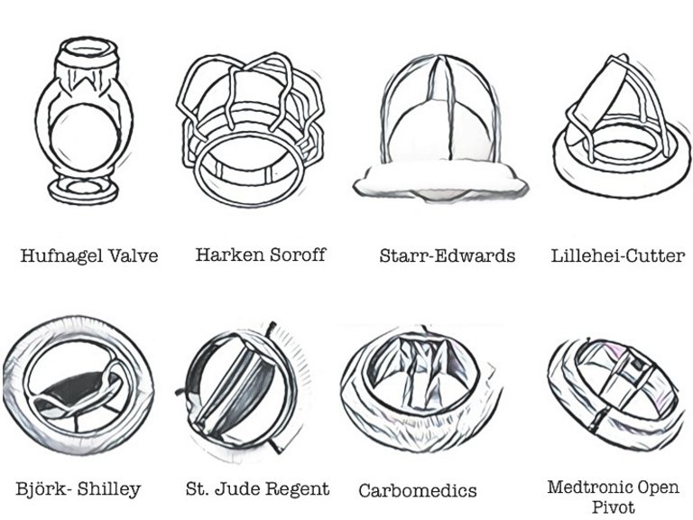
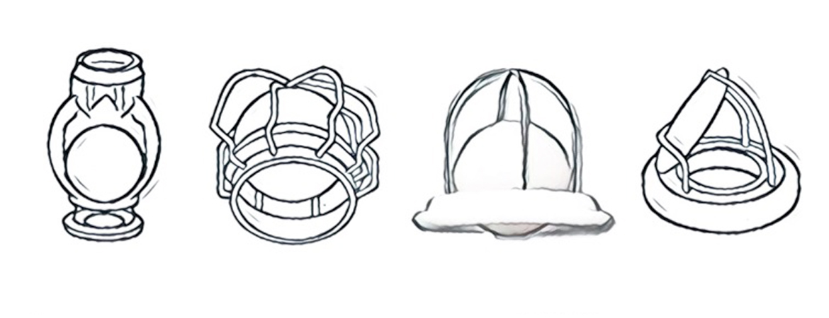
After more than 50 years of evolution, mechanical valve replacement represents an optimal treatment for patients with heart valve disease. Mortality decreased progressively and no differences in term of prognosis have been described when comparing mechanical with biological valves [16–17]. Figure 1 shows the evolution of mechanical heart valve prostheses.
Figure 1: Mechanical heart valve evolution. From Hufnagel heart valve to the the current bileaflet prostheses.
But what can we expect in the current era from this old tool? Could innovation in valvular heart therapies ­alter the role of mechanical valves? Could mechanical valves benefit from new anticoagulation strategies?
Studies in animals showed that dabigatran was effective in preventing valve thrombosis and was associated with reduced mortality after mitral valve surgery. These encouraging data have not yet translated into human practice [18]. New materials could be less thrombogenic and patient selection could be redefined accordingly, but there is not yet sufficient evidence to change the standard anticoagulation management.
As a matter of fact, today most patients prefer to receive a tissue valve, to avoid anticoagulation, and the age threshold is continually reduced in guidelines, ranging between 60 and 65 years.

Reducing anticoagulation-related events: the advent of biological valves

The evolution of biological tissue valves is a mix of ­biochemistry, mechanical engineering and biology. A tissue valve provides some clear advantages in terms of biocompatibility, with concerns related to its durability.
The history of tissue valves originated from evidence of the haemodynamic and biological advantages of cadaveric homografts, first implanted in the aortic position by Donald Ross in 1962 [19]. His effort was largely based on the premise that “our entire physical makeup and body structures represent the end result of millions of years of evolutionary development”[20], and the assumption that no prosthetic valve can replicate such perfection.
Since homograft cadaveric valves were difficult to collect and preserve, the next step was to use xenografts – valves collected from animals. The first generation of biological valves was substantially consisted of porcine valves, the valves most similar to human ones. Several new issues were debated. How can these xenografts be preserved and how made immunologically inactive? What is the haemodynamics of non-human valves and their durability after implantation?
Tissue valve engineering began with the use of formalin to sterilise and fix the fresh xenograft tissue. This technique was complicated by collagen breakdown, with risk of early cusp calcification and occurrence of fibrosis with a big shortfall in expected valve dura­bility. Remembering the origin, Carpentier wrote some years later: “It became obvious that the future of tissue valves would depend upon the development of methods of preparation capable of preventing inflammatory cell reaction, and penetration into the tissue” [21]. Therefore, he suggested the use of glutaraldehyde for the chemical treatment of porcine valves [22]. Creating cross-links in collagen molecules, this treatment protected the leaflets from denaturation and made the tissue immunological inactive due to ­antigen modification. Anticalcification treatment changed the history of tissue valves, increasing the expected durability. Moreover, in 1966 Carpentier began to mount the whole porcine valve into a stent, obtaining a proper three dimensional space relationship between the leaflets and simplifying the implantation technique. From the haemodynamic standpoint, a central flow was achieved but further analyses revealed an important pressure drop attributed to several factors, such as the restriction of leaflet opening caused by the stent, the stiffness of the fixed geometry imposed by the pig’s anatomy and the presence of artificial commissures. The roles played by haemodynamic factors, mechanical stress and biological response in structural valve failure led to a growing interest in alternative strategies and new materials to improve outcomes [12].

Increasing the durability and improving haemodynamics: from porcine to peri­cardial

As postulated by Carpentier, an understanding of the chemical properties of biological tissue led to continuous and intensive research into the creation of a bioprosthesis that would provide longer freedom from structural deterioration.
Bovine pericardium was identified as a promising ­alternative tissue source for producing artificial leaflets, because of its histological and physical characteristics in terms of thickness, pliability, abundance and wide availability [23]. In 1971, Ionescu in Leeds started the production and implantation of pericardial heart valves. The concept was to create a completely “man-made” prosthesis, to optimise the anatomical configuration and avoid the fixed geometry of an animal valve. Bovine pericardium treated with glutaraldehyde was mounted on Delirin flexible stent, in order to achieve a synchronous opening of the three leaflets (Ionescu-Shiley valve). In vitro haemodynamic studies showed more symmetrical opening than with the porcine ones. Despite the first enthusiasm, after 5 years of follow-up the first cases of structural valve deterioration (SVD) were detected. Analysis of the explanted valves revealed that the leaflets were torn by movements within the stent. The mode of failure was very unfortunate, and led to sudden severe aortic regurgitation, ­occasionally fatal. The technique of suturing the pericardium onto the stent was modified, such that it was sewn in the outermost part, in order to reduce impingement. Moreover, different types of stent were introduced; these were more flexible and thinner, with stress reduction in the commissural site, and allowed supra-anular implantation so that larger prostheses could be used [23].
To improve durability, after 1980 most prostheses were developed by treating the leaflets with zero- or low-pressure fixation. The goal of these methods was to maintain a more normal morphology of the leaflets. Several antimineralisation methods were invented by different companies to obtain durable leaflets, and characterised the continuous evolution of bio­logical valves [6].
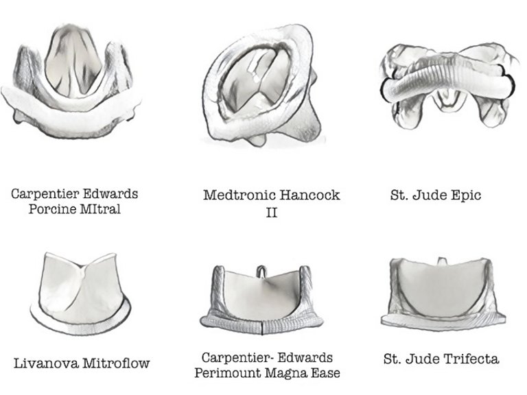
Figure 2 summarises schematically biological prostheses for the mitral and aortic positions.
Figure 2: Biological heart valves evolution. In the first line mitral prostheses, porcine (Carpentier Edwards Porcine) and pericardial (Hancock II and Epic). In the second line 3 rd  generation of aortic prostheses.

Patient-prosthesis mismatch: how to manage it by use of different prostheses

Firstly reported by Rahimtoola in 1978, patient-prosthesis mismatch (PPM) represents an important issue in current practice [24]. Patients with valves with an EOA too small for their body size develop PPM and are at higher risk of postoperative mortality, reduced mass regression and limited functional benefit.
The negative impact of PPM on patient prognosis after aortic valve replacement has been reported in several studies showing an increased risk of mortality and SVD [25]. As previously mentioned, a totally supra-anular valve implantation technique was proposed: the third generation of bioprostheses (St. Jude Trifecta, Sorin Mitroflow, Carpentier-Edwards Perimount Magna) were designed to achieve a larger EOA through modification of the stent architecture, but here surgical technique plays a major role. Surgeons should be aware of the consequences of implantation of a valve too small for the patient, and avoid it.
Various alternative solutions have been suggested to overcome to the issue of PPM. Stentless valves were introduced by Tirone David in 1988. They are xenograft, both porcine and pericardial, without any stent or sewing cuff, and represent the extreme of the continuous reduction in valvular stent dimensions. Providing a large valve orifice and improved haemodynamics, they could theoretically induce a greater reduction of ventricular mass and avoid PPM. These promising results are balanced by a more difficult and time-consuming implantation, which requires specific skills in aortic root surgery.
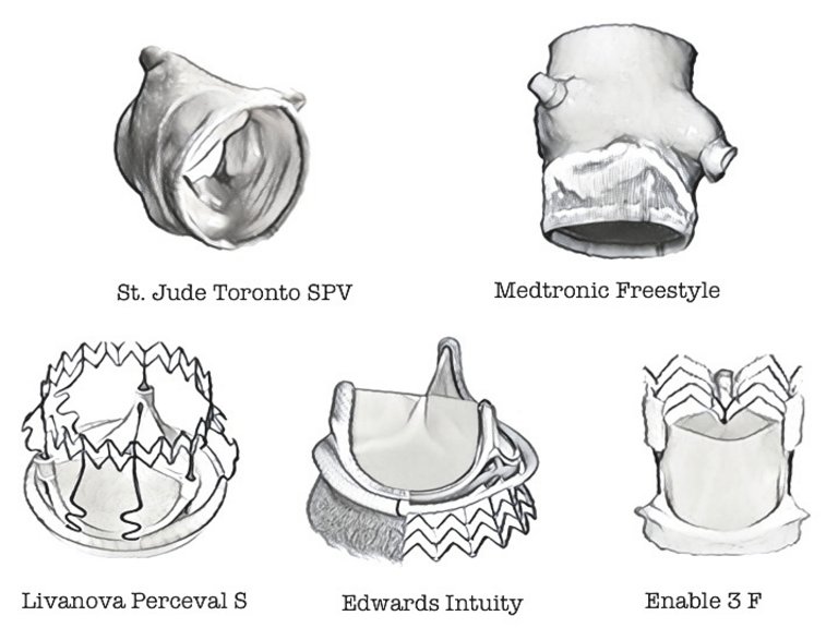
Initial experience with stentless prostheses revealed a high rate of perioperative aortic regurgitation due to a discrepancy between the valve annulus and the native sino-tubular junction. Complete root replacement was thus encouraged and new prostheses were developed, such as the complete porcine root (fig. 3). The great enthusiasm for these valves culminated in the late 1990s, and faded because no superiority over stented valves was detected in long-term studies [26]. The stentless technology made a big contribution to the next wave of valve technology evolution. Both sutureless and trans­catheter valves were designed on the foundation of stentless bioprostheses, and furthermore, several new antimineralisation strategies and the use of equine pericardium (3F aortic bioprosthesis) were developed during the evolution of stentless valves.
Figure 3: Stentless and sutureless aortic prostheses.

A route to less invasiveness: the role of sutureless aortic valves

During the 1990s, minimally invasive cardiac surgery was rapidly developing [27]. This concept brings at least two benefits: a reduction in surgical access in order to minimise surgical trauma and wound complication and a reduction in cross-clamp and cardiopulmonary bypass time. Moreover, several datasets showed that the prevalence of frail patients with heart valve disease, and aortic stenosis in particular, was progressively increasing [28].
Three valves were introduced: the Livanova Perceval S, the Edwards Intuity and the Enable 3F (fig. 3). The aim was to reduce surgical time by avoiding the use of sutures to fix the valve to the annulus as a result of a new stent configuration, which can expand and thus anchor the valve in the right position [29]. The stent characteristics depend on the properties of nitinol, which has memory of shape and becomes flexible according to the temperature. Although several studies showed optimal results with sutureless valve implantation ­instead of an increased risk of complete atrioventricular block and residual paravalvular leaks, the use of 3F has been discontinued owing to late valve migration. Haemodynamic features were comparable to those of stentless prostheses, but long-term durability is still unknown [30].
Although sutureless aortic valves were initially intended for intermediate-high risk patients, the rapid ­development of transcatheter valve technologies profoundly affected the course of their evolution. Their current role is still to be clarified, but several conditions, such as small aortic root, multiple valve surgery, or use as a facilitating tool in minimal invasive aortic valve surgery could represent fields of application.

The last step of the evolution: transcatheter valve procedures bring surgery back to the time of “closed-heart” procedures

Transcatheter valve interventions are the most advanced development in cardiac surgery and were initially introduced as the ideal solution to the new epidemiological scenario of a large number of untreated elderly and high-risk patients with aortic stenosis. Transcatheter valves take advantage of decades of valve ­evolution to deliver surgical grade interventions involving ­miniaturised instruments (catheter-based devices) by an endovascular approach, without the need of cardiopulmonary bypass and cardioplegia [31].
Percutaneous mitral valve commissurotomy was the first surgical treatment converted into a transcatheter procedure. Its development is a model in the field. Initially, balloon valvuloplasty was restricted to high-risk and inoperable patients; more recently, balloon valvuloplasty became the gold standard treatment for all comers, and surgery is performed only in patients with anatomical contraindications to transcatheter treatment. The first implantation of a transcatheter valve into a human was performed in 2000, when Bonhoeffer implanted a pulmonary transcatheter valve [32]. Two years later, this approach was translated to the aortic position by Cribier, with worldwide clinical resonance [33].
When read together, the first report of mechanical heart valve implantation by Starr [9] and the Cribier’s first transcatheter aortic valve implantation (TAVI) report [33] have many similarities. Just as Starr treated end-stage patients, so TAVI was introduced as a “last ­resort” solution. The same enthusiasm and the same passion of a cardiac surgeon in 1960 and a cardiologist about 40 years later characterise the two papers. This parallel demonstrates how the evolution represents a continuous cycle of different solutions to treatment of the same pathology, with continuously new technologies. Each step is a fundamental contribution to know­ledge and fosters further developments. Many problems in this process can be avoided by reading and digesting the history of previous mistakes.
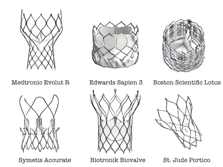
The development of percutaneous heart valves brought together the evolution of bio-valves, stents and delivery catheter design. In order to permit endovascular releasing, the prosthesis should be crimped, with a decrease in dimensions of more than three folds without any damage to the leaflet. Two types of stents were developed: the stainless steel baloon-expandable stent and the self-expanding nitinol ones (fig. 4).
Figure 4: Transcatheter aortic valve implantation devices currently in use.
Since the beginning of this technology several issues have been identified as potential limiting factors: paravalvular leakage, a high rate of vascular complications, risk of neurological events and complete atrioventricular block.
The Cribier-Edwards (previously PVT) balloon-expandable valve (Edwards Lifesciences) was the first transcatheter aortic prosthesis (2002). It consisted initially of equine pericardium and a stainless-steel frame. In order to improve sealing, a polyethylene terephthalate fabric skirt was introduced; this modification represented the first Edwards SAPIEN model (2006) [34]. ­Owing to the high profile of the delivery system, several patients were treated via a transapical approach. The SAPIEN XT (2009) valve was then designed with a lower-profile tubular cobalt-chromium stent that made it possible to downsize it to reduce peripheral ­access complications and increase the use of the transfemoral approach. The last development of the SAPIEN valve is the SAPIEN 3 (2013), in which an additional outer skirt was added to increase sealing and an ­expandable 14/16 F sheet was designed to minimise femoral invasiveness. All the valves were treated with an anticalcification process involving glutaraldehyde fixation and phospholipid extraction, and a new “mild-heat” treatment that removes unstable glutaraldehyde molecules was introduced.
The prototype of self-expandable valves is represented by the Medtronic Corevalve (2005). This consists of peri­cardial leaflets mounted on a nitinol frame. The first-generation leaflets were made of bovine peri­cardium, but a switch to porcine pericardium, together with the use of a more flared outflow design, allowed the development of a lower profile device. The evolution of the Corevalve resulted in the EVOLUT R. Several improvements made this device repositionable, resheathable and recapturable, and the height and ­diameter of the delivery system were reduced. Recently the Evolut PRO device was approved by the US Food and Drug Administration. New features include an outer wrap that adds surface area contact between the valve and the native aortic annulus to improve valve sealing.
Innovation profoundly changed the clinical use of TAVI. In contrast to the early stages, when its use was limited to high risk and inoperable patients, inter­mediate-risk patients are currently treated since recent data showed that TAVI is a non-inferior, and sometime superior, alternative to surgery in the short term. The design of TAVI valves gives them optimal haemodynamic results [35], which might support the clinical superiority, particularly in patients at risk of PPM.
The transcatheter approach is also used in the treatment of atrioventricular valve diseases, most of all in repair procedures. Recently, transcatheter mitral valve implantation (TMVI) became an option for patients with degenerated bioprosthesis or with recurrence of mitral regurgitation after ring annuloplasty. Although TMVI presents a number of challenges as a result of the native anatomy, its feasibility in high-risk patients with functional and degenerative valve disease has been recently reported [36].
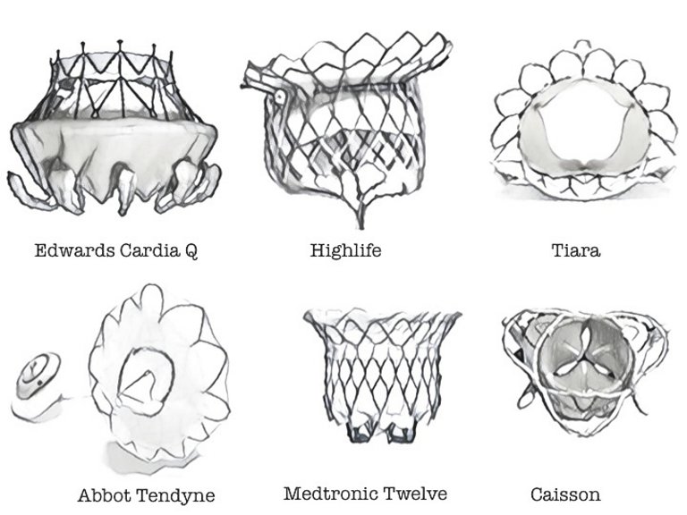
Several devices (fig. 5) have been introduced, but the procedure is still technically demanding and the ­patient’s anatomy is still a controversial issue for feasibility. Risk of left ventricular outflow tract obstruction, optimal fixation to the native mitral annulus and ­access nowadays represent the greatest challenges in TMVI procedures [37].
Figure 5: Transcatheter mitral valve implantation prostheses.
Whether to repair or replace the mitral valve was for a long time a matter of debate in the surgical context. Similarly, we could expect that, once a reliable replacement device becomes available, most operators would abandon repair. However, with time and experience, valve repair could come back as an option to limit the drawbacks of a permanent implant in the mitral position (Starr and Edwards said the same in the 1960s, a prediction which turned out to be true today for surgery).

How the “new” valves are changing the “old” valves

“Valve-in-valve” procedures have been recently introduced and rapidly became the treatment of choice, in order to avoid surgical reoperation, for patients who experienced the limited durability of bioprostheses. The anatomical characteristics and the size of the previously implanted valve represent the major limiting factors for the implantation of a TAVI valve in valve. This new therapeutic scenario created a new need for ­bioprosthesis design to provide a more efficient “re-valving” procedure in the future and provide patients and surgeons an ad-hoc platform from which to expand indications for tissue valves in the aortic position, and possibly also in the mitral position, to a population younger than 60 years of age.
The INSPIRIS valve (Edwards Lifescience) was developed as a new class of surgical valves. The Cobalt-chromium stent has an area of possible expansion that gives the valve the capability to be enlarged in the case of a future valve-in-valve procedure. Moreover, the bovine pericardial tissue is transformed by means of a novel integrity preservation technology that eliminates free aldehyde molecules while protecting and preserving the tissue [38]. The COMMENCE Trial to evaluate the results of this promising technology, also the in mitral and pulmonary positions, is ongoing.

Beyond the present: tissue-engineered heart valves

All the devices described exhibit a lack in remodelling and growth capability. This concept has led to the ­development of innovative valve substitutes called regenerative valves or tissue-engineered valves (TEHVs). This novel approach is based on various tissue engineering technologies that provide an alternative crimp­able valve replacement device thought to be a ­definitive solution, also for younger and paediatric ­patients [39].
A TEHV would be a living organ, capable of responding and growing like the native valve. The immune response plays a special role in regulating remodelling after implantation. This technology aims to become the most advanced means to improve valve durability [40].
Experience with TEHVs is still preclinical and, even if transcatheter implantation is successfully performed in animal models, the way the device could interact with a calcified annulus must be clarified, before it can be translated into clinical practice [39, 40].

What can we expect from new-era ­prostheses and new-era physicians?

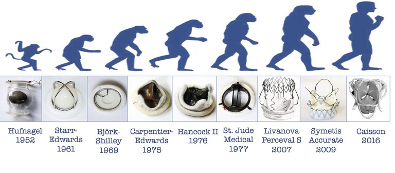
The long process of evolution of heart valves demonstrates how innovation induces changes in practice and contributes to better patient treatment. Different subcategories of patient and new challenges have been overcome during almost one century of cardiovascular interventions. And the story is not finished yet (fig. 6).
Figure 6: Evolutionary steps in heart valve technology. Images courtesy of Prof. von Segesser [4].
The latest evolution of transcatheter therapies has ­induced a revolution in clinical practice, moving the view from “operator-related” to “patient-related”. The concept of “heart team” was introduced in order to ­define which patient could benefit from a particular treatment. Cardiac surgeons, cardiologists, anaesthesiologists, imaging physicians and dedicated nurses, started to work in cooperation to build a new environment of cardiovascular medicine, focused on patient-centred care. Creating new competences and new ­evidence nowadays represents the main goal of our profession. In this ever evolving landscape, looking back into history will pave the way to the future.
Abbreviation list:
SVD = structural valve deterioration
EOA = effective orifice area
PPM = patient-prosthesis mismatch
TAVI = transcatheter aortic valve implantation
THV = transcatheter heart valve
TMVI = transcatheter mitral valve implantation
F. Nietlispach is a consultant for Abbott and Edwards Lifesciences. F. Maisano is consultant surgeon for Abbott, Medtronic and St. Jude. The other authors have no conflict of interest to declare.
Correspondence:
Francesco Maisano MD, FESC
University Heart Center
Rämistrasse 100
CH-8091 Zürich
francesco.maisano[at]usz.ch
 1 Rehn L. On operating cardiac injuries and cardiac suturing. Arch Klein Chir. 1897;55:315.
 2 Tuffier T. État actuel de la chirurgie intrathoracique. Trans Int Congr Med. 1913 7; Surgery 1914;2:249.
 3 Hufnagel CA. Aortic plastic valvular prosthesis. Bull Georgetown Univ Med Center. 1951;5:128–30.
 4 Von Segesser LK. The development of open valve surgery. In: Thoracic and Cardiovascular Surgery. From the magin mountain to rocket science. European Association for Cardio-Thoracic Sugery; 2010. p. 178–89.
 5 Gott VL, Alejo DE, Cemeron DE. Mechanical heart valve: 50 years of evolutions. Ann Thorac Surg. 2003;76:S2230–9.
 6 Chiariello L. Storia della Cardiochirurgia. In: Trattato di chirurgia Cardiaca. Universo Edition; 2016. p. 3–22.
 7 Harken DE, Soro HS, Taylor WJ, et al. Partial and complete prostheses in aortic insufficiency. J Thorac Cardiovasc Surg. 1960;40:744–62.
 8 Braunwald NS, Cooper T, Morrow AG. Clinical and experimental replacement of the mitral valve. Experience with the use of a flexible polyurethane prosthesis. in: Merendino K.A. (Ed.) Prosthetic Valves for Cardiac Surgery. Springfield, Ill.: Charles C Thomas; 1961. p. 307–31.
 9 Starr A, Edwards ML. Mitral replacement: Clinical experience with a ball-valve prosthesis. Ann Surg. 1961;154:726–40.
10 Gödje OL1, Fischlein T, Adelhard K, Nollert G, Klinner W, Reichart B. Thirty-year results of Starr-Edwards prostheses in the aortic and mitral position. Ann Thorac Surg. 1997;63:613–9.
11 Gudbjartsson T, Absi T, Aranki S. Mitral valve replacement. In: Cohn L (ed.). Cardiac Surgery in the adult. New York: McGraw-Hill; p. 1032–68.
12 Chaikof EL. The Development of Prosthetic Heart Valves – Lessons in Form and Function. N Engl J Med. 2007;357:1368–71.
13 Lindblum D, Rodriguez L, Bjork VO. Mechanical failure of the Bjork-Shiley valve: updated follow-up and considerations on prophylactic replacement. J Thorac Cardiovasc Surg. 1989;97:95–7.
14 Nicoloff DM, Emery RW, Arom KV, et al. Clinical and hemodynamic results with the St. Jude Medical cardiac valve prosthesis. J Thorac Cardiovasc Surg. 1982;82:674–83.
15 Czer LS, Chaux A, Matloff JM, et al: Ten-year experience with the St Jude Medical valve for primary valve replacement. J Thorac ­Cardiovasc Surg. 1990;100:44; discussion 54.
16 Palatianos GM, Laczkovics AM, Simon P, et al. Multicentered European study on safety and effectiveness of the On-X prosthetic heart valve: intermediate follow-up. Ann Thorac Surg. 2007;83:40–6.
17 Vahanian A, Alfieri O, Andreotti F, et al. Guidelines on the management of valvular heart disease (version 2012). Eur Heart J. 2012;33:2451–96.
18 Puskas J, Gerdisch M, Nichols D, et al. Reduced anticoagulation after mechanical aortic valve replacement: interim results from the prospective randomized on-X valve anticoagulation clinical trial randomized Food and Drug Administration investigational device exemption trial. J Thorac Cardiovasc Surg. 2014;147:1202–10.
19 Ross DN. Homograft replacement of the aortic valve. Lancet. 1962;2:487.
20 Ross DN. Evolution of the Homograft Valve. Ann Thorac Surg. 1995;59:565–7.
21 Spencer FC: Intellectual creativity in thoracic surgeons. J Thorac
Cardiovasc Surg. 1983;86:168.
22 Carpetier A, Lemaigre G, Robert L, et al. Biological factors affecting long term results of valvular heterograft. Jour Thorac Card Vasc Surg. 1969;28:467–83.
23 Ioneuscu MI. The begining. In: The pericardial heart valve. Society for Cardiothoracic Surgery in Great Britain and Ireland; 2014; p. 1–35.
24 Rahimtoola SH. The problem of valve prosthesis-patient ­mismatch. Circulation. 1978;58:20–4.
25 Flameng W, Rega F, Vercalsteren M, et al. Antimineralization treatment and patient-prosthesis mismatch are major determinants of the onset and incidence of structural valve degeneration in bioprosthetic heart valves. J Thorac Cardiovasc Surg. 2014;147:1219–24.
26 Desai ND, Merin O, Cohen GN, et al. Long-term results of aortic valve replacement with the St. Jude Toronto stentless porcine valve. Ann Thorac Surg. 2004;78:2076–83.
27 Cosgrove DM 3rd, Sabik JF. Minimally invasive approach for aortic valve operations. Ann Thorac Surg. 1996;62:596.
28 Osnabrugge RL, Mylotte D, Head SJ, et al. Aortic stenosis in the ­elderly: disease prevalence and number of candidates for transcatheter aortic valve replacement: a meta-analysis and modeling study. J Am Coll Cardiol. 2013;62:1002–12.
29 Comendador JM, Castano M, Gualis J et al. Sutureless aortic ­bioprosthesis. Interact Card Vasc Surg. 2018:1–8.
30 Shrestha M, Fischlein T, Meuris B, et al. European multicentre ­experience with the sutureless perceval valve: clinical and haemodynamic outcomes up to 5 years in over 700 patients. Eur J Cardiothorac Surg. 2016;49:234–41.
31 Maisano F, Nietlispach F. The future of valve for percutanues ­insertion. Eur Heart J. 2014:35:1569–74.
32 Bonhoeffer P, Boudjemline Y, Saliba Z et al. Percutaneous replacement of pulmonary valve in a right- ventricle to pulmonary-artery prosthetic conduit with valve dysfunction. Lancet. 2000;21:9239.
33 Cribier A, Eltchaninoff H, Bash A et al. Percutaneous transcatheter implantation of an aortic valve prosthesis for calcific aortic stenosis: First human case description. Circulation. 2002;106:3006–8.
34 Abdel-Wahab M, Jose J, Richardt G. Transfemoral TAVI devices: ­design overview and clinical outcomes. EuroIntervention. 2015;11(Suppl W):W114–8.
35 Reardon MJ, Van Mieghem NM, Popma JJ. Surgical or Transcatheter Aortic-Valve Replacement in Intermediate-Risk Patients. N Engl J Med. 2017;376:1321–31.
36 Maisano F, Kuwata S, Nietlispach, et al. Transcatheter mitral valve repair and replacement.J Cardiovasc Med (Hagerstown). 2017;17 (Suppl 1):e134-e140.
37 Taramasso M, Kuwata S, Rodriguez Cetina Biefer H, et al. Percutaneous mitral valve repair and replacement: complementary or competitive techniques? EuroIntervention. 2016;12(Y):Y97–Y101.
38 Flameng W, et al. A randomized assessment of an advanced tissue preservation technology in the juvenile sheep model. J Thorac ­Cardiovasc Surg. 2015;149:340–5.
39 Fioretta ES, Dijkmann PE, Emmert MY, et al. The future of heart valve replacement: recent developments and translational challenges fo heart valve tissue engineering. J Tissue Eng Regen Med. 2016 Sep 30. doi: 10.1002/term.2326. [Epub ahead of print]
40 Emmert MY, Fioretta ES, Hoerstrup SP. Translational Challenges in Cardiovascular Tissue Engineering. Cardiovasc Transl Res 2017. doi: 10.1007/s12265-017-9728-2.
With the comment function, we offer space for an open and critical exchange of expertise. We publish comments as long as they comply with our guidelines.