Sinus venosus and unroofed coronary sinus defects: two cases diagnosed and treated in adulthood

Case Report
Issue
2019/06
DOI:
https://doi.org/10.4414/cvm.2019.02074
Cardiovasc Med. 2019;22:w02074

Affiliations
a Division of Cardiology, University Hospitals of Geneva, Switzerland.
b Division of Cardiovascular Surgery, University Hospitals of Geneva, Switzerland.
c Division of Radiology, University Hospitals of Geneva, Switzerland.
d Division of Pneumology, University Hospitals of Geneva, Switzerland.

Published on 17.12.2019

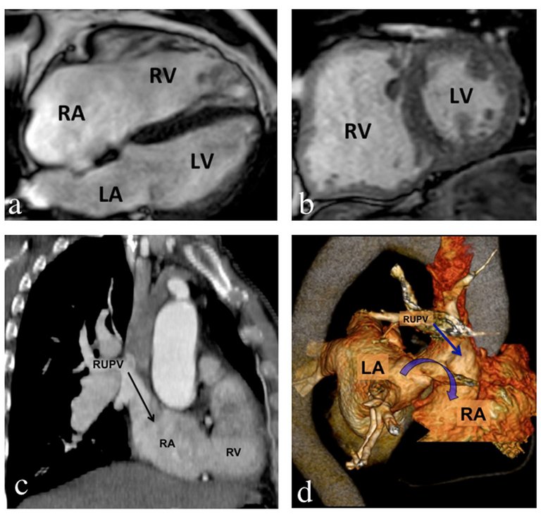
Interatrial communications are the most frequent congenital heart malformation in adulthood. We report typical examples of sinus venosus and unroofed coronary sinus defects associated with anomalous pulmonary venous return.
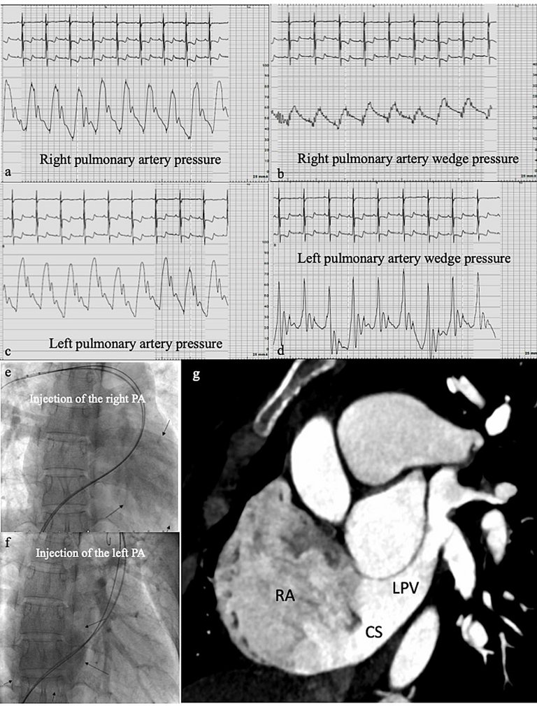
Introduction

Interatrial communications are the most frequent congenital heart malformations diagnosed in adulthood, after bicuspid aortic valve. Four different types of atrial defect (AD) are described. Ostium secundum atrial septal defect (ASD) is the most frequent form and accounts for approximately 75% of all ADs, the ostium primum form represents 15%, the sinus venosus form 5–10% and the coronary sinus form less than 1% [1]. When the left-to-right shunt is significant, right heart cavities enlarge in response to volume overload and atrial arrhythmias may develop, most commonly after the fourth decade of life. Patients with a left-to-right shunt are at risk of developing pulmonary artery hypertension (PAH). In the Euro Heart Survey of adults with congenital heart disease, 34% of the patients with an open AD had systolic pulmonary pressure ≥40 mm Hg, whereas this rate decreased to 12% in patients with a closed AD [2]. We report the cases of two patients diagnosed in adulthood with typical forms of superior sinus venosus and unroofed coronary sinus defects.

Case reports

Case 1: sinus venosus defect

A 68-year-old patient without cardiovascular risk factors was admitted for progressive dyspnoea. Physical examination revealed signs of right heart failure. The ECG showed atrial flutter with right bundle branch block, without signs of myocardial ischaemia. Transthoracic echocardiography (TTE) showed a normal-sized left ventricle with severe dysfunction and a left ventricular ejection fraction (LVEF) of 30–35%. The right heart cavities were severely dilated. Cardiac magnetic resonance imaging (MRI) (fig. 1A and B) and computed tomography (CT) (fig. 1D) confirmed the dilatation of the right atrium and the right ventricle, presumably due to a superior sinus venosus defect (curved arrow) and partial anomalous pulmonary venous return of the right upper pulmonary vein (RUPV) to the right atrium.
Figure 1
Sinous venosus defect. A,B. Cardiac magnetic resonance imaging showing dilatation of the right atrium (RA) and right ventricle (RV). C. Right upper pulmonary venous (RUPV) return into the RA. D. Reconstructed images of computed tomography scan showing a superior sinus venosus defect (curved arrow) with anomalous RUPV return into the RA.
LA = left atrium; LV = left ventricle
Following medical treatment of the heart failure, the patient underwent left and right cardiac catheterisation, which showed a normal left ventricular end-diastolic pressure (10 mm Hg) and a significant left-to-right shunt with a pulmonary flow (Qp) / systemic flow (Qs) ratio of 2.4, and excluded pulmonary hypertension (systolic pulmonary artery pressure [PAP] 43 mm Hg, mean PAP 22 mm Hg, without significant differences between the two pulmonary arteries). Because of the anatomy, our preferred technique for correction of sinus venosus defects is the Warden procedure [3]. Surgery in this case consisted of transecting the superior vena cava (SVC) above the anomalous pulmonary vein, tunnelling the lower SVC and anomalous RUPV through the AD to the left atrium using a pericardial patch, and anastomosing the SVC to the right atrial appendage. The patient had a favourable outcome. At day 7, TTE showed normalisation of the right heart cavities size and LVEF. At day 10, the patient was discharged home. At 18-month follow-up, the patient reported a favourable evolution without limitation of physical activity. TTE showed normalisation of the volume of the right cavities, associated with normal left and right ventricular function without indirect signs of pulmonary hypertension.

Case 2: unroofed coronary sinus defect

A 39-year-old patient, with a history of surgical patent foramen ovale (PFO) closure 20 years ago in Mongolia (no report available), complained of progressive dyspnoea on exertion associated with palpitations. Physical examination was considered normal apart from a systolic heart murmur at the left sternal border without radiation. The ECG showed regular sinus rhythm with right axis and negative T waves on lead II, III, aVF, and V1-V6. TTE and transoesophageal echocardiography (TOE) showed a dilated and hypertrophied right ventricle with diminished systolic function (tricuspid annular plane systolic excursion [TAPSE] 9 mm) and a LVEF of 65%. There was a high probability of pulmonary hypertension, (dilatation of the main pulmonary artery, mild pulmonary regurgitation and tricuspid maximal velocity of 405 cm/sec). There was also a supra-mitral membrane in the infero-posterior wall of the left atrium, with a dilated coronary sinus to which the left pulmonary veins were connected, compatible with a partially unroofed coronary sinus defect.
Right and left heart catheterisation with injection of the left and right pulmonary arteries confirmed the anomalous left pulmonary venous return to the coronary sinus (fig. 2A–F) and showed severe pulmonary hypertension with partial reversibility with oxygen and no additional decrease under nitric oxide: PAP (systolic/diastolic-mean) was 82/39-57 mm Hg at rest, 72/27-48 mm Hg with oxygen and 72/30-49 mm Hg with nitric oxide. The wedge pressure was 24 mm Hg in the right pulmonary artery and was normal in the left pulmonary artery. Qp/Qs was 1.63, the systolic aortic pressure was 107 mm Hg and the systolic PAP was 72 mm Hg (ratio 67%) (table 1). CT (fig. 2G) also showed the left pulmonary venous return to the coronary sinus. Despite the elevated PAP, the patient underwent surgical repair, which is the only option to correct a coronary sinus defect.
Figure 2
Coronary sinus defect. A. Right pulmonary artery pressure (scale on 100 mm Hg), systolic/diastolic-mean (s/d-m) 82/39-57 mm Hg. B. Right pulmonary arterial wedge pressure. (scale on 40 mm Hg), V wave-A wave/mean 21-24/21. C. Left pulmonary artery pressure. (scale on 100 mm Hg) s/d-m 76/36-52 mm Hg. D. Left artery wedge pressure is low owing to the venous return of the two left pulmonary veins into the coronary sinus, which communicates with the right atrium. (scale on 40 mm Hg), V wave-A wave/mean 26-12/10. E. Right pulmonary (PA) artery injection showing the left heart cavities (black arrows). F. Left PA injection showing the right heart cavities (black arrows), and an abnormal pulmonary venous return. G. Computed tomography image showing the left pulmonary venous return to the coronary sinus.
CS = coronary sinus; LPV = left pulmonary vein; RA = right atrium
Table 1
Right and left heart catheterisation of patient 2.
 BaselineWith oxygenWith nitric oxide
Systemic pressure
(mm Hg; s/d-m)
107/60-83
Systolic PAP (mm Hg)Right: 82
Left: 76
7272
Diastolic PAP (mm Hg)Right: 39
Left: 36
2730
Mean PAP (mm Hg)Right: 57
Left: 52
48*49*
Wedge (mm Hg)Right: 21
Left: 10
LVEDP (mm Hg)15
Qp/Qs1.63
Systemic cardiac output (l/min)3.74
Pulmonary cardiac output (l/min)6.10
TPG36
PVR (WU)(57-21)/6.1=5.9
SVR (WU)(83-2)/3.74= 21.6
LVEDP = left ventricular end diastolic pressure; PAP = pulmonary arterial pressure; PVR= pulmonary vascular resistance; Qp = pulmonary blood flow; Qs = systemic blood flow; s/d-m = systolic/diastolic-mean; SVR = systemic vascular resistance; TPG = trans pulmonary pressure gradient
* similar results for right and left pulmonary arteries
No left SVC was noted on surgical inspection. The supra-mitral membrane was resected, and the coronary sinus was surgically completely unroofed to allow better drainage of the pulmonary veins into the left atrium. Finally, the dilated coronary sinus ostium (residual septal defect) was closed using a pericardial patch. During surgery a suture was observed at the site of the PFO closure 20 years before. At day 4 after surgery, TTE showed normal left ventricular size and function, with persistent dilatation of the right ventricle and moderately impaired function. The estimate of PAP was in favour of persistent severe pulmonary hypertension (tricuspid maximum velocity 379 cm/sec).
After being lost to follow up for 2 years, the patient came back and reported clinical improvement. TTE showed normalisation of the size of the right heart cavities, as well as of right and left ventricular function, but persistence of indirect signs of pulmonary hypertension (tricuspid maximum velocity 308 cm/sec). Cardiac catheterisation was repeated and revealed persistence of severe precapillary pulmonary hypertension (mean PAP 46 mm Hg, wedge pressure 9 mm Hg, vascular pulmonary resistance 8.8 WU) with no vasoreactivity to nitric oxide. Upfront PAH-specific double therapy with an oral endothelin receptor antagonist and a phosphodiesterase 5 inhibitor was introduced, in accordance with the 2015 European guidelines on the diagnosis and treatment of pulmonary hypertension.
One year after introduction of PAH treatment, a new right heart catheterisation showed persistent mild precapillary pulmonary hypertension, with an important reduction of pulmonary vascular resistance and mean PAP (mean PAP 33 mm Hg, diastolic PAP 21 mm Hg, wedge pressure 6 mm Hg, cardiac output 6.4 l/min and vascular pulmonary resistance 4.2 WU).
N-terminal pro B-type natriuretic peptide was 220 ng/l (reference range <300) and cardiac MRI showed a biventricular functional improvement (LVEF 64 vs 60% 2 years previously; RVEF 54 vs 48% 2 years previously).
Therefore, the association of surgery and PAH-specific double therapy permitted normalisation of size and function of the right heart cavities with clinical improvement.

Discussion

We describe two typical cases of the less common interatrial communication forms, which were diagnosed in adulthood. The sinus venosus form – accounting for 5–10% of ADs – is a communication between the two atria localised between the junction of the vena cava and the left atrium, with an unroofing of the pulmonary veins at the level of the AD. The majority of cases involve a superior sinus venosus. In 85% of cases we observe an anomalous pulmonary venous return (usually a right upper pulmonary vein) associated with this pathology. The unroofed coronary sinus defect is the rarest form of interatrial communication, accounting for <1% of all ADs. It is frequently associated with a persistent left superior vena cava, and anomalous pulmonary venous return has been described in this form of AD [3].
Right cardiac chamber dilatation, even in asymptomatic patients, should arouse suspicion of an AD. TTE can help with identification and localisation of the defect, but sinus venosus and unroofed coronary sinus ADs can easily be missed on TTE and the investigations should include TOE and/or cardiac MRI to assess the anatomy of the interatrial septum and the drainage of the pulmonary veins. Of note, MRI can also be used to calculate the shunt and TTE with agitated saline might expose a right-to-left shunt or a wash-out phenomenon in the presence of a left-to-right shunt.
The treatment depends on the type of AD and the importance of the associated left-to-right shunting. Ostium secundum ASDs are increasingly treated via a percutaneous approach, whereas the ostium primum, sinus venosus and coronary sinus ADs are still surgically treated (mini-sternotomy, thoracotomy) [3].
The goals of surgical therapy are to unload the dilated right ventricle by closing the AD, and redirecting the systemic venous drainage to the right atrium and the pulmonary venous drainage to the left atrium. The lesions can be repaired with standard cardiopulmonary bypass techniques and systemic venous cannulation.
In most cases of sinus venosus defect, two options exist, depending on the level of the most superior anomalous right pulmonary vein: either a double patch (tunnelling the pulmonary veins to the left atrium with a patch and augmenting if necessary the junction of the SVC and right atrium to avoid stenosis from the tunnelling) versus a Warden procedure as described above. Because the double patch repair places the sinus node at risk (although surgical series have not shown an increased risk of pacemaker implantation associated with this technique) [4], and because the systemic or pulmonary venous pathways are at risk of stenosis, the Warden procedure is our preferred technique. For coronary sinus defects, direct suturing or patch insertion for extensive unroofed cases allows rerouting of the coronary sinus to the right atrium and separation of the venous and systemic circulation. In cases with partially anomalous left pulmonary venous return to the coronary sinus, as in our case, two options exist: (1) transection of anomalous pulmonary veins and anastomosis to the left atrial appendage, and correction of any defects in the coronary sinus wall; (2) unroofing of the coronary sinus and closure of the coronary sinus ostium by primary suture and/or patch insertion (admittedly leaving the coronary venous blood to drain into the left atrium). We preferred the second option, as patch repair of an unroofed coronary sinus is at risk of creating an obstruction in the coronary sinus if the patch is too small, or a supramitral stenosis if the patch is too large.
Importantly, haemodynamically significant ADs must be corrected as soon as they are diagnosed in adults and before school age in children. It has, however, been shown that even elderly patients will benefit from AD closure, with regression of right cardiac chamber dilatation, even though diastolic dysfunction increases with age and is frequently seen in patients over the age of 60 years [5]. In the elderly, diastolic dysfunction may increase the risk of acute left ventricular failure and pulmonary oedema after AD closure owing to the elimination of a low impedance leak [6]. Therefore, the assessment of left atrial pressure is recommended for patients at risk of diastolic dysfunction, such as hypertensive or elderly patients. A balloon occlusion test is recommended when the mean left atrial pressure exceeds 15 mm Hg in order to identify a restrictive physiology. In patients with an increase of ≥10 mm Hg in left atrial pressure during the occlusion test, diuretic therapy may be used before AD closure; preconditioning using an angiotensin converting-enzyme inhibitor and diuretic for 4 weeks may be an alternative strategy in patients with left-sided filling pressures over 25 mm Hg during transient balloon occlusion of the AD [7]. Incomplete or fenestrated AD closure (fenestrated ASD occluder when a transcatheter approach is used) may be required in patients at risk of pulmonary congestion despite pretreatment [5].

Conclusion

In conclusion, these two reported cases confirm that after multidisciplinary and specialised assessment patients’ age may not be a limiting factor for AD closure, and that post-surgical outcome can be good, with regression of right heart cavity size and improvement of cardiac function. However, close follow-up by centres expert in adult congenital heart disease is essential to optimise management after closure and even more so in the context of pulmonary hypertension.
Dr S. Noble, MD, Structural Cardiology Unit, Cardiology Division, Department of Medicine, University Hospitals of Geneva, Rue Gabrielle-Perret-Gentil 4, CH-1211 Geneva, stephane.noble[at]hcuge.ch
1 Marelli AJ, Mackie AS, Ionescu-Ittu R, Rahme E, Pilote L. Congenital heart disease in the general population: changing prevalence and age distribution. Circulation. 2007;115(2):163–72. doi:. http://dx.doi.org/10.1161/CIRCULATIONAHA.106.627224 PubMed
2 Gatzoulis MA, Alonso-Gonzalez R, Beghetti M. Pulmonary arterial hypertension in paediatric and adult patients with congenital heart disease. Eur Respir Rev. 2009;18(113):154–61. doi:. http://dx.doi.org/10.1183/09059180.00003309 PubMed
3 Rao PS, Harris AD. Recent advances in managing septal defects: atrial septal defects. F1000 Res. 2017;6:2042. doi:. http://dx.doi.org/10.12688/f1000research.11844.1 PubMed
4 Walker RE, Mayer JE, Alexander ME, Walsh EP, Berul CI. Paucity of sinus node dysfunction following repair of sinus venosus defects in children. Am J Cardiol. 2001;87(10):1223–6, A8. doi:. http://dx.doi.org/10.1016/S0002-9149(01)01504-1 PubMed
5 Noble S, Ibrahim R. Percutaneous atrial septal defect closure in patients with left ventricle failure or pulmonary hypertension. Prog Pediatr Cardiol. 2012;34(2):109–12. doi:. http://dx.doi.org/10.1016/j.ppedcard.2012.08.009
6 Tomai F, Gaspardone A, Papa M, Polisca P. Acute left ventricular failure after transcatheter closure of a secundum atrial septal defect in a patient with coronary artery disease: a critical reappraisal. Catheter Cardiovasc Interv. 2002;55(1):97–9. doi:. http://dx.doi.org/10.1002/ccd.10068 PubMed
7 Gruner C, Akkaya E, Kretschmar O, Roffi M, Corti R, Jenni R, et al.Pharmacologic preconditioning therapy prior to atrial septal defect closure in patients at high risk for acute pulmonary edema. J Interv Cardiol. 2012;25(5):505–12. doi:. http://dx.doi.org/10.1111/j.1540-8183.2012.00747.x PubMed